Plastic surgery Health care - covered by insurance Beauty care – paid by the patient
Gynaecology Prenatal diagnosis
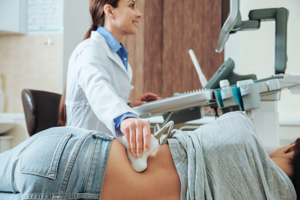
Medical genetics Urology Orthopaedics X-ray site Rehabilitation
Office hours Restaurant
Not sure? Contact us through our form HERE.

Kidney, ureter surgery due to stones, conventional approach
What does this procedure entail?
The procedure is based on the removal of a urinary stone from the renal pelvis/calyx or ureter. This method accesses the kidney via an incision in the lumbar region – in the ureter, the incision is sometimes performed through the lower abdomen depending on the position of the stone. These methods are today rather exceptional – conditions not allowing for other approaches or connection with another surgery.
What are the alternatives to this procedure?
- Laparoscopic rendevouz
- LERV – Lithotrypsy via extracorporeal shock wave – stone disintegration by targeted high-energy pulses
- Percutaneous extraction (PEK) from the kidney or from the upper part of the ureter – the stone is removed endoscopically through a hole in the lumbar region
- Ureteroscopy – the stone is removed from the ureter endoscopically through the bladder
- Stent insertion (temporarily inserted silicone tube)
What should I expect before this procedure?
If you regularly use Anopyrin, Clopidogrel, Warfarin, etc., you must tell your doctor. These medications can be associated with increased risk of bleeding during and after the surgery if they are not stopped in time. In some cases, it is necessary to replace these drugs with injection preparations. It is necessary to consult your general practitioner or cardiologist about this approach. At the request of the referring physician, you will have to schedule preoperative examinations to assess your overall condition, including laboratory and instrumental tests. If the report does not preclude the indicated procedure, you will be admitted to the hospital. If you don’t undergo a preoperative examination or it is incomplete, you will not be able to have the surgery as scheduled. You will be admitted by a nurse and a member of the medical team will complete your examination and assess your fitness for the surgery.
You will be asked not to eat or drink for at least 6 hours before the surgery!
In the evening before the surgery, you will receive medication from an anaesthesiologist to calm you down so that you sleep well.
Remember to inform your physician about the following possible facts before the surgery:
- artificial heart valve
- coronary artery stent
- pacemaker
- artificial joint
- artificial vascular graft
- neurosurgical bypass
- other implanted foreign body
- use of the following prescription drugs: Acylpyrin, Anopyrin, Aspirin, Godasal, Clopidogrel, Plavix, Kardegic, Aspegic, Micristin, Ibustrin,Ticlid, Tagren, Ipaton Apo-Tic, Plavix, Persantin, Curantyl, Anturan, Aggrenox, Vessel due F.
- drug and other allergies
- any abnormalities or eventualities
It is NECESSARY to inform the physician about your use of drugs affecting blood clotting before your admission for the procedure.
What will happen during the surgery?
You will normally receive an injection or oral antibiotics before the procedure, but any allergies must be checked first.
The procedure is performed exclusively under general anaesthesia.
The surgery is sometimes started with cystoscopy (examination of the bladder using a telescopic instrument inserted into the urethra), where a ureteral catheter or stent is inserted into the renal pelvis. The kidney is then accessed from an incision in the lumbar region, it can be guided to the ureter in the lower abdomen and the stone is then removed by opening the renal pelvis or the ureter. After the surgery, a urinary catheter is inserted into the bladder (to collect urine), a ureteral tube or stent can be left in the ureter (improves the drainage of the renal excretory system after the surgery). Exceptionally, a nephrostomy drainage tube is inserted into the renal excretory system (draining urine from the kidney through the skin); a tubular drain is inserted into the kidney/ureter (draining blood and tissue secretions and possibly urine leaking from the surgical wound). These drains are brought to the surface in the area of the hip or lower abdomen.
This procedure takes between 60 and 120 minutes.
Sometimes a short X-ray examination (fluoroscopy) is used during the surgery to specify the position of the stones.
What will happen immediately after the procedure?
You will wake up at the intensive care unit equipped for the continued monitoring of patients immediately after the procedure. The surgeon will inform you about the course of the procedure. You will still have high levels of anaesthetics in your blood at that time, so you may not remember this conversation.
Patients usually have an infusion access point into a vein in their arm; if necessary, a monitoring/infusion port (a tube inserted into the vein to monitor blood pressure or administer drugs and nutrition) will be inserted into a vein (jugular vein) in the supraclavicular region (the area between the collarbone and neck).
Nutrition is provided in an intravenous manner shortly after the surgery. You will receive a liquid and mushy diet for the next 2 to 3 days. This procedure is necessary for the proper restoration of gastrointestinal function. Non-compliance of the patient is usually associated with severe abdominal pain, vomiting with the risk of suffocation and may require surgical revision (further surgery in case of complications).
Physical rehabilitation is very important to prevent complications in the postoperative period. You will first exercise on the bed. As soon as your medical condition allows, you will be allowed to sit down and then stand up. After that, you will be able to walk slowly and carefully around the room, initially accompanied by medical staff. Pay close attention to eventual dizziness, uncontrollable weakness, and to gait stability. Otherwise immediately inform medical personnel or anyone in the vicinity.
The drain from the wound is removed after morning secretion ceases, usually after 3-5 days. In the case of nephrostomy drains, it is about 7 days. The urinary catheter is removed after sufficient mobilisation, and the stent is usually only removed subsequently in an outpatient manner. Sutures will be removed before you are discharged from the hospital. The average hospitalisation is 9 days.
Once you are fully conscious, you should:
- ask if the planned outcome was achieved
- inform the medical staff about any problems
- ask what you can and cannot do
- ask all the questions you have for the healthcare professionals and members of the medical team.
- remember (and understand) why the surgery was performed, how it turned out, and what will follow
What are the postoperative risks or complications?
Common (10% of procedures of this type)
- Introduction of bladder urinary catheter and wound drain
- Insertion of stent / nephrostomy drain and their subsequent removal
- The stent may be associated with more frequent urination urges, a feeling of discomfort or mild pain in the bladder area (especially when urinating). Sometimes, blood may appear in the urine.
- Bulging at the incision site due to damage to the nerves of the abdominal wall (interruption of muscle innervation when approaching the kidney/ureter)
Occasional (2-10% of procedures of this type)
- Bleeding requiring further surgery or transfusion
- Risk of additional or relapsing stones
Rare (may occur in 2% of procedures of this type)
- Cardiovascular or anaesthesia-related complications that may require a longer stay at the intensive care unit (lung infections, pulmonary embolism, heart attack, deep vein thrombosis, etc.
- Injuries to nearby surrounding structures (blood vessels, spleen, liver, lungs, pancreas and intestines) or the kidney/ureter itself requiring more extensive surgery or planned revision, rarely the removal of the kidney
- Long-term urine drainage due to poor wound healing in the renal pelvis
- Scarring/narrowing of the ureter due to surgery requiring further surgery
- Infection, pain or hernia in the wound requiring further therapy
- Possibility that all stones will not be removed
- A need for further intervention to remove stones
- Recurrent kidney or bladder infections.
Hospital infections
- MRSA colonisation (0.9% – 1 of 110)
- Intestinal infection by clostridium difficile (0.01% – 1 of 10,000)
- MRSA blood infection (0.02% – 1 of 5,000)
- Hospital infection rates may be higher in high-risk patients, such as in cases requiring long-term drainage, after a previous infection, after prolonged hospitalisation or after multiple hospitalisations.
What should I expect when I return home?
When you are discharged from the hospital, you should:
- Get recommendations on recovery at home
- Ask when you can return to normal activities such as work, exercise, driving, housework
- Get a contact number for further questions after returning home or in case of trouble
- Ask about the date and place your sutures will be removed and subsequent check-ups will be performed (hospital or your doctor)
- Make sure you are aware of the reason, course and outcome of the surgery, the results of examinations or the removal of tissues or organs.
At your departure from the hospital (sometimes several days later), you will receive a hospitalisation report. The document contains important information about your hospital stay, your surgery and recommended follow-up. If you need to call your attending physician or visit the hospital for any reason, take this document with you so that the physician knows the details of your treatment. This is especially important if you need to consult another doctor or longer after discharge.
What else should I watch out for?
It is recommended that you wear elastic stockings for 2-3 weeks after the discharge from the hospital.
Uncomplicated skin incisions will heal in about 14 days. After that, you will be able to take a regular shower. The healing of internal structures takes 6 to 12 weeks, so it is necessary to maintain a resting regime even after discharge from the hospital. It usually takes 8-16 weeks until you are able to return to work. Talk about this with the doctor who treated you in the hospital, or at least with your general practitioner.
Many patients complain about persistent episodes of pain (pulling, pressure, pulsing) in the lumbar region, which can last for several months. If the kidney/ureter access incision is in the lumbar region, the wound may arch due to the interruption of the nerves supplying the muscles of the abdominal cavity – it is not a hernia. This condition can be improved by strengthening the muscles of the abdominal wall with exercise (only after full healing!).
Important information?
In case of fever, redness, pulsation or secretion at the site of the surgical wound, contact your attending physician. Tell your doctor if you have any other problems associated with the procedure.
Date and place of your check-up at the outpatient urological office or at the GP will be determined upon discharge (usually 6-8 weeks after the surgery). Stent removal, if it was inserted, will be scheduled as well.
Further follow-up at the outpatient urological office is a usual practice.
We cooperate with health insurance companies
in the Czech Republic